
ISPprofawardees

Immunohistochemistry (IHC)
Immunohistochemistry (IHC) is a laboratory technique used to detect specific proteins in tissue sections by using antibodies that bind to those proteins. It combines principles of immunology (antibody-antigen interactions) with histology (study of tissue structure).
IHC is widely used in research, diagnostics, and pathology to study tissue architecture, disease states, and protein localization.
How IHC Works
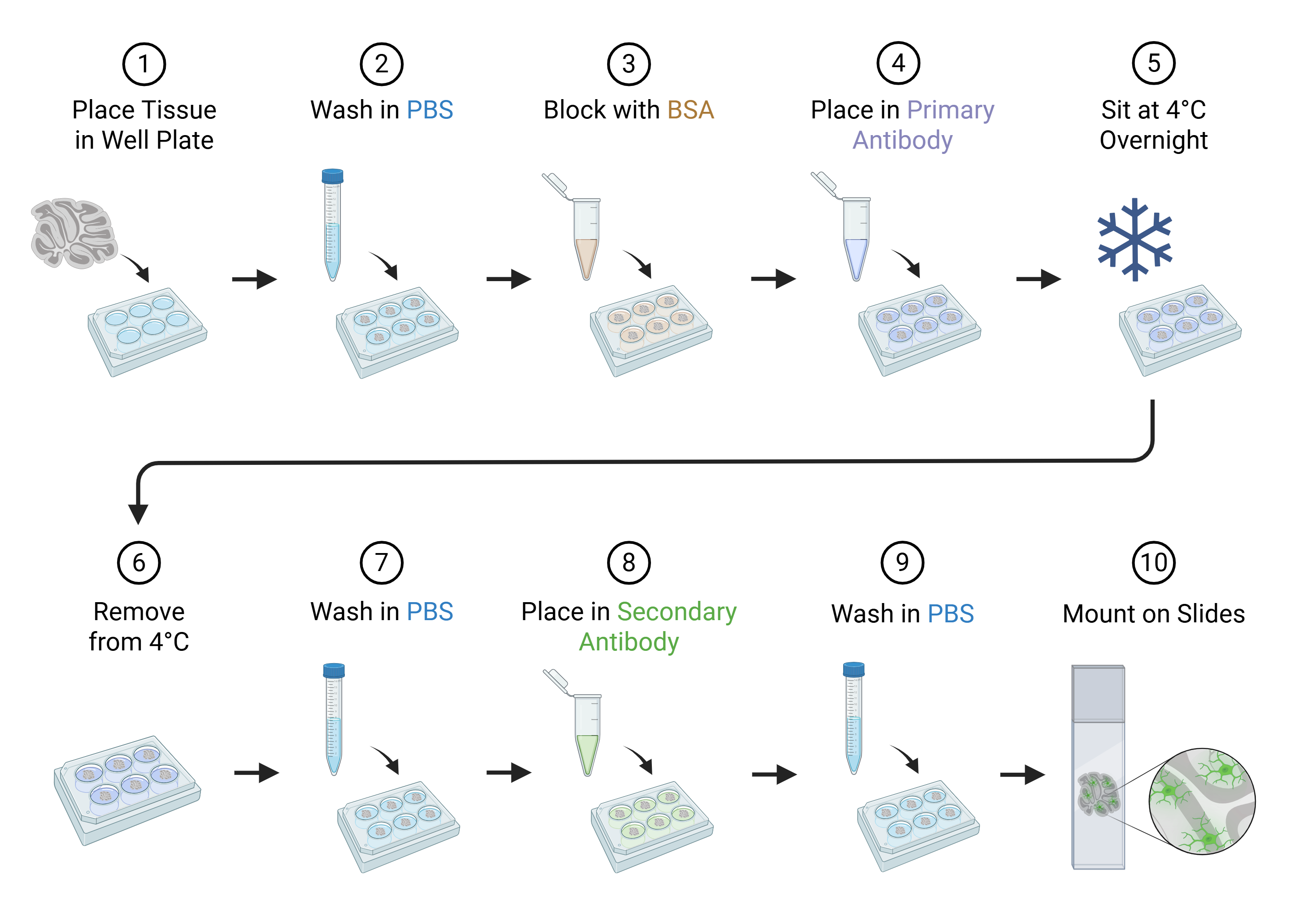
Sample Preparation:
Tissue is collected and preserved, usually by formalin fixation and embedding in paraffin.
Sections of the tissue (thin slices) are mounted on glass slides.
Antigen Retrieval:
Fixation can mask protein sites (antigens).
Methods like heat-induced epitope retrieval (HIER) or enzyme treatment are used to unmask these antigens.
Blocking:
Non-specific binding sites are blocked to reduce background staining using serum or protein solutions.
Primary Antibody Application:
A specific primary antibody binds to the target protein (antigen) in the tissue.
Secondary Antibody Application:
A secondary antibody, which recognizes the primary antibody, is applied.
It is usually linked to an enzyme (like HRP) or a fluorescent dye.
Detection:
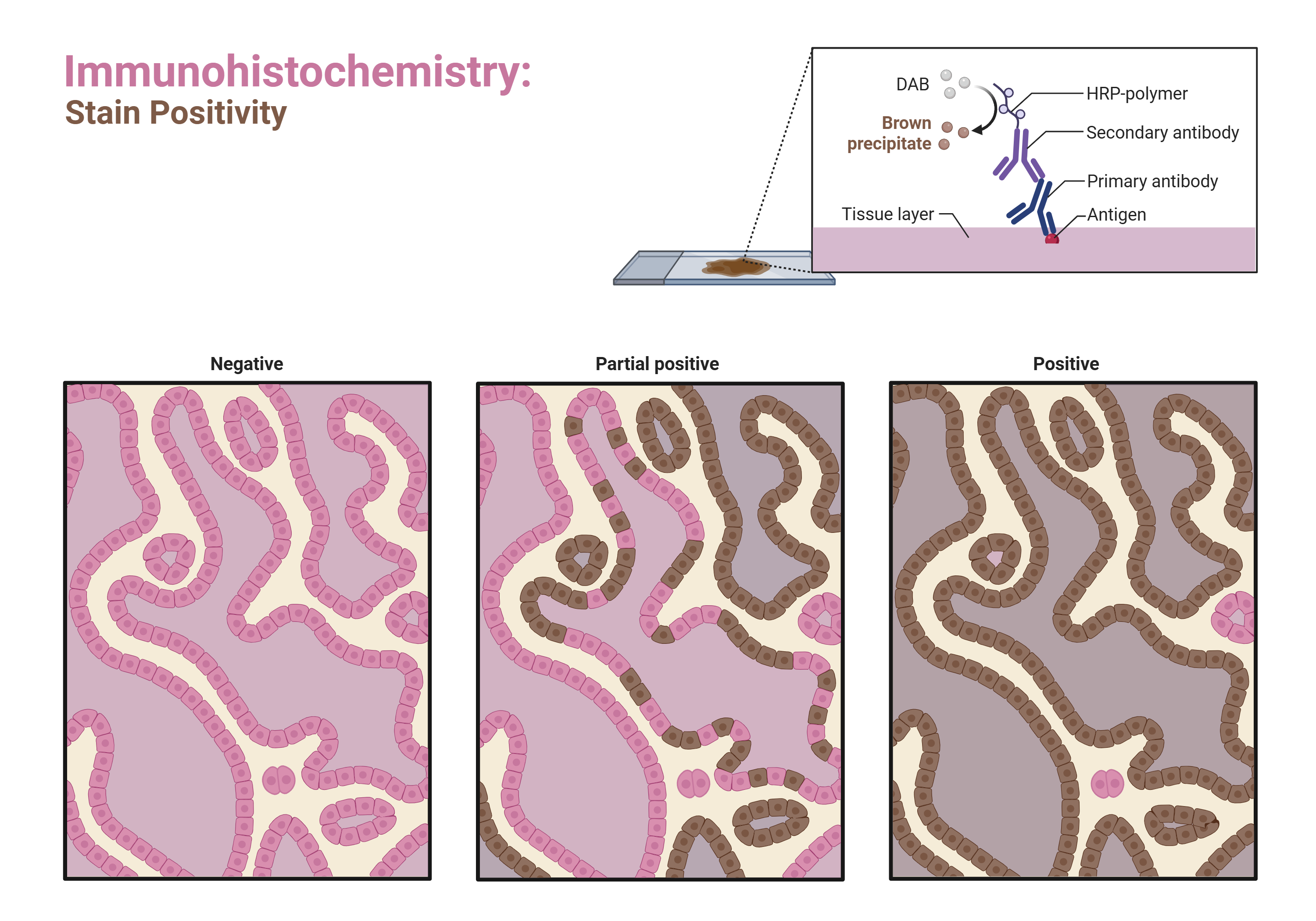
For enzyme-linked antibodies: a substrate (e.g., DAB) produces a colored precipitate at the site of the antigen.
For fluorescent antibodies: the signal is visualized under a fluorescence microscope.
Counterstaining and Mounting:
A counterstain (like hematoxylin) highlights tissue structure.
Slides are then examined under a microscope.
Professional Award Recipients in Immunohistochemistry
The field of Immunohistochemistry is advanced by pioneering scientists and medical professionals whose work leads to critical discoveries in disease mechanisms, diagnostic accuracy, and therapeutic development. Professional Award Recipients in IHC are recognized for their significant contributions, which may include:
-
Developing novel antibody clones with superior specificity.
-
Innovating antigen retrieval techniques that revolutionized tissue staining.
-
Creating standardized scoring systems for consistent diagnostic interpretation.
-
Pioneering multiplex IHC methods for complex protein analysis.
Honoring these Professional Award Recipients is essential, as their expertise drives the entire field forward, ensuring that IHC remains a gold standard in diagnostic pathology and biomedical research.
Applications of IHC
Cancer Diagnostics: Detects tumor markers, e.g., HER2 in breast cancer or PD-L1 in immunotherapy planning.
Research: Maps protein expression in tissues.
Pathogen Detection: Identifies viral or bacterial antigens in tissue samples.
Neuroscience: Visualizes proteins in brain tissues.
Advantages
High specificity due to antibody-antigen interaction.
Provides spatial context within the tissue.
Can differentiate between cell types and subtypes.
Limitations
Requires high-quality antibodies.
Subject to variability in staining and interpretation.
May have background noise if blocking is insufficient.
Types of IHC
Direct IHC: Primary antibody is directly labeled. Quick but less sensitive.
Indirect IHC: Secondary antibody amplifies the signal. More common in research and diagnostics.
Fluorescent IHC (IF): Uses fluorophores instead of enzymes.
Multiplex IHC: Detects multiple antigens simultaneously on the same tissue section.